
Frontotemporal Dementia: The Forgotten Frontier of Neurodegeneration
The Hidden Face of Dementia: When Personality Changes Before Memory

Introduction: The Many Faces of FTD
Frontotemporal dementia (FTD) is one of the most challenging and misunderstood neurodegenerative conditions of our time. Once thought to be rare, it’s now recognized as a leading cause of dementia in people under 65—striking at a stage of life when work, family, and identity are often at their fullest. What makes FTD so devastating isn’t only how it changes language, behavior, or personality—it’s how it quietly reshapes the brain’s very circuits for empathy, connection, and emotion, often leaving loved ones feeling as though the person they knew is slowly slipping away.
To understand how medicine came to recognize and untangle this complex condition, we have to journey back—through the early observations of Arnold Pick in the 19th century, the decades of confusion that followed, and the modern genetic and molecular discoveries that are finally beginning to illuminate its causes and point toward hope for the future.
The History and Nomenclature of FTD
Pick’s Disease: The Beginning
The story begins with Arnold Pick, a Czech neurologist who, in the 1890s, described patients who slowly lost the ability to speak and reason while their memory and awareness initially remained intact. Autopsies revealed distinct areas of brain shrinkage—particularly in the frontal and temporal lobes.
Alois Alzheimer later identified the abnormal microscopic inclusions in these patients, which became known as “Pick bodies.” For decades, the term Pick’s disease referred broadly to all frontotemporal degenerations.
Refining the Diagnosis: The Mid-20th Century Pioneers
Throughout the 1900s, neuropathologists such as Delay, Brion, Escourolle, Malamud, and Constantinidis began to notice that not all “Pick’s disease” cases were alike. Some had classic Pick bodies, others did not. Some presented with language loss, others with personality changes.
Their work slowly dismantled the idea of a single “Pick’s disease” and laid the groundwork for recognizing frontotemporal lobar degeneration (FTLD) as a family of related disorders rather than one disease.
The Splintering of Pick’s Disease
By the late 20th century, advances in microscopy and molecular biology revealed that these disorders had distinct underlying proteins—tau, TDP-43, and FUS—accumulating in neurons and glia. What had once been a single disease name now fractured into several subtypes: behavioral variant FTD (bvFTD), primary progressive aphasia (PPA), corticobasal degeneration (CBD), progressive supranuclear palsy (PSP), and others.
This shift marked the birth of the modern concept of frontotemporal dementia.
Corticobasal Degeneration: Another Piece of the Puzzle
In the 1960s and 70s, researchers like Rebeiz, Gibbs, Riley, and Lang described patients with asymmetric rigidity, apraxia, and cortical sensory loss. The pathology—abnormal accumulations of tau protein—became known as corticobasal degeneration (CBD). Though its symptoms often begin as movement problems, CBD shares molecular features with FTD, underscoring how overlapping tauopathies can express as either cognitive or motor syndromes.
Frontal Lobar Dementia and the Birth of FTD
In the 1980s and 1990s, scientists such as Gustafson, Brun, Neary, Snowden, Bowen, and Mann formalized new clinical entities. They coined terms like frontal lobe dementia, dementia of frontal type, and eventually frontotemporal dementia (FTD).
Neary and Snowden’s work at the University of Manchester was particularly influential, defining diagnostic criteria for the behavioral variant of FTD (bvFTD)—a syndrome characterized by social disinhibition, apathy, compulsivity, and loss of empathy.
Primary Progressive Aphasia (PPA): A New Language of Dementia
In 1982, neurologist Marsel Mesulam described six patients with slowly progressive loss of language but preserved memory and reasoning. This became known as primary progressive aphasia (PPA)—a linguistic counterpart to bvFTD.
Over the next decades, researchers including Norman Geschwind, John Hodges, Julie Snowden, Karalyn Patterson, and Marilu Gorno-Tempini helped classify PPA into three main subtypes:
Semantic variant (svPPA): Loss of word meaning
Nonfluent/agrammatic variant (nfvPPA): Effortful, halting speech
Logopenic variant (lvPPA): Difficulty retrieving words and repeating phrases
Each subtype maps onto distinct regions of the left temporal or frontal lobes, highlighting how specific neural networks underpin language and meaning.
From UCLA to UCSF: The Modern Research Era
In recent decades, major research centers such as UCLA and UCSF have transformed our understanding of FTD. Through advanced neuroimaging, genetic mapping, and brain banking, these institutions have linked specific mutations and proteins to distinct clinical patterns. Their work has also revealed that the emotional and social brain—particularly the frontoinsular and anterior cingulate cortices—is central to what makes us human, and tragically, what FTD most directly erodes.
The Biological Revolution
The Genetic Breakthroughs
The modern era of FTD research began when familial forms of the disease were discovered. Roughly 40–50% of FTD cases now show a clear hereditary component.
The main genetic culprits include:
MAPT: Mutation in the tau protein gene, leading to abnormal microtubule stabilization.
GRN: Progranulin deficiency, disrupting cellular repair and inflammation control.
C9ORF72: A hexanucleotide repeat expansion linked to both FTD and ALS.
TARDBP, FUS, VCP, CHMP2B, and SQSTM1: Rarer mutations affecting protein handling and degradation.
These discoveries reframed FTD as a molecular network disorder—a breakdown in how brain cells fold, traffic, and recycle essential proteins.
Neuropathology: The Proteins That Define the Disease
Neuropathologists now classify frontotemporal lobar degeneration (FTLD) by the dominant misfolded protein:
FTLD-tau: Includes Pick’s disease, PSP, and CBD.
FTLD-TDP: Involving TDP-43 inclusions, often seen in FTD-ALS and PPA.
FTLD-FUS: A rare form involving the FUS protein.
These proteins accumulate in selective networks—especially the anterior temporal, orbitofrontal, and insular cortices—leading to degeneration of specialized neurons such as von Economo neurons, which are critical for social awareness and empathy.
FTD Today: From Behavior to Biomarkers
A Spectrum, Not a Single Disease
Frontotemporal dementia is best understood as a family of disorders. The same gene mutation may produce different clinical symptoms, and the same behavioral pattern may arise from different molecular pathologies.
Clinically, FTD can mimic psychiatric illness, Alzheimer’s disease, or even motor neuron disease. Yet its hallmark remains the erosion of social and emotional cognition—the networks that make interpersonal life possible.
Diagnosis and Research Frontiers
Modern diagnosis combines clinical history, neuropsychological testing, MRI or PET imaging, and increasingly, blood or CSF biomarkers. Advanced tools like tau-PET imaging and genetic testing are helping clinicians predict the underlying pathology long before autopsy.
Artificial intelligence, machine learning, and connectome mapping are also being used to detect subtle changes in speech, facial emotion, and social behavior that precede clinical diagnosis.
How Frontotemporal Dementia Is Diagnosed
Clinical History and Observation
Diagnosis begins with a detailed history and behavioral observation, often with input from family members or close friends who notice changes first.
Clinicians look for:
Personality change (loss of empathy, disinhibition, apathy)
Changes in eating or compulsive behaviors
Social withdrawal or poor judgment
Speech or language difficulty
Emotional blunting or loss of insight
Key diagnostic principle:
In FTD, behavior and language deteriorate first, while memory may stay intact early on — unlike in Alzheimer’s disease.
Neurological and Cognitive Examination
A neurologist will perform a comprehensive neurological and mental status exam, checking:
Speech fluency, grammar, and word comprehension
Executive functions (planning, reasoning, mental flexibility)
Emotional regulation and social awareness
Motor and eye movement abnormalities (may suggest PSP or CBD variants)
Neuropsychological Testing
Formal neuropsychological assessment helps differentiate Frontotemporal Dementia (FTD) from other dementias and psychiatric conditions.
Here are the most commonly used domains and tests:
Executive Function
Example Tests: Stroop Test, Trail Making Test (Part B), Wisconsin Card Sorting Test
What They Reveal: Difficulty shifting mental sets, reduced cognitive flexibility, and poor impulse control
Language
Example Tests: Boston Naming Test, Western Aphasia Battery, Pyramids and Palm Trees Test
What They Reveal: Identifies Primary Progressive Aphasia (PPA) variants — semantic, nonfluent, or logopenic
Social Cognition
Example Tests: Faux Pas Test, Reading the Mind in the Eyes Test, Facial Emotion Recognition
What They Reveal: Measures empathy, emotional understanding, and theory of mind — often impaired in behavioral variant FTD (bvFTD)
Memory
Example Tests: Rey Auditory Verbal Learning Test, Wechsler Memory Scale
What They Reveal: Memory is often relatively preserved in early bvFTD, helping distinguish it from Alzheimer’s disease
Visuospatial Skills
Example Tests: Rey–Osterrieth Complex Figure, Clock Drawing
What They Reveal: Visuospatial abilities are usually intact early in FTD — another key feature differentiating it from Alzheimer’s
4. Neuroimaging
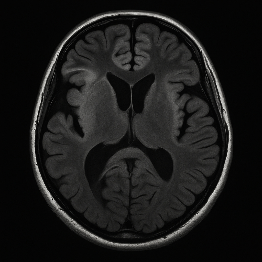
MRI (Magnetic Resonance Imaging)
The most common imaging test for FTD.
Findings:Frontal and/or anterior temporal lobe atrophy
Asymmetry depending on subtype (right = behavioral changes; left = language loss)
“Knife-edge” atrophy of gyri in advanced stages
FDG-PET (Fluorodeoxyglucose Positron Emission Tomography)
Shows hypometabolism (low glucose use) in frontal and temporal regions even before visible shrinkage on MRI.
Amyloid and Tau PET
Amyloid PET: Typically negative in FTD (helps rule out Alzheimer’s).
Tau PET: Emerging as a biomarker for certain tauopathies (e.g., PSP, CBD).
DTI (Diffusion Tensor Imaging) / fMRI
Advanced research tools to visualize white-matter disconnection and network dysfunction.
Biomarkers and Laboratory Testing
Cerebrospinal Fluid (CSF) Analysis
Used mainly to rule out Alzheimer’s and other causes:
Low Aβ42 and high tau → Alzheimer’s pattern
Normal or non-Alzheimer’s pattern → supports FTD diagnosis
Research biomarkers (under development):Neurofilament light chain (NfL) – elevated in FTD
Progranulin levels – low in GRN mutation carriers
Blood Biomarkers (Emerging)
Plasma NfL, GFAP, and tau fragments are being studied as less invasive screening tools.
Genetic Testing
Because up to half of FTD cases are familial, genetic counseling and testing are often offered—especially if symptoms start before age 65 or there’s a family history of dementia, ALS, or psychiatric disease.
Key genes tested:MAPT (microtubule-associated protein tau)
GRN (progranulin)
C9ORF72 (repeat expansion linked to ALS)
FUS, TARDBP, VCP, CHMP2B, SQSTM1 (rare)
Additional and Supportive Tests
EEG: Usually normal or nonspecific (helps rule out seizures or Creutzfeldt-Jakob disease).
Neuropsychiatric evaluation: Screens for depression, bipolar disorder, or psychosis mimicking FTD.
Functional assessments: Activities of daily living (ADLs), caregiver questionnaires, and social behavior scales.
Differential Diagnosis Checklist
Clinicians always compare FTD against other disorders that can present similarly:
Alzheimer’s disease – memory first, not behavior
Major depression or bipolar disorder – mood changes precede cognition
Schizophrenia or late-onset psychosis – psychotic symptoms dominate
Chronic traumatic encephalopathy (CTE) – history of repetitive head trauma
Vascular dementia – stepwise progression, vascular lesions on MRI
Metabolic / infectious / autoimmune encephalopathies – reversible causes
Key Diagnostic Insight
No single test “proves” FTD — diagnosis comes from pattern recognition:
clinical behavior + cognitive testing + neuroimaging + (when indicated) genetic or biomarker confirmation.Early identification is vital, as it allows families to plan care, participate in research trials, and consider targeted treatments as they emerge.
The Current Treatment Landscape
Unlike Alzheimer’s disease, there is no FDA-approved drug specifically for FTD.
That said, doctors can manage symptoms, support caregivers, and in some cases slow functional decline by combining medications, behavioral strategies, and lifestyle interventions.
A. Medications for Symptom Management
Serotonergic Agents (SSRIs)
Selective serotonin reuptake inhibitors (SSRIs) such as sertraline, citalopram, fluoxetine, or paroxetine are often first-line for:
Disinhibition
Compulsive behaviors
Emotional blunting or apathy
Irritability
Why they help: the serotonergic system is significantly depleted in FTD, particularly in the orbitofrontal and anterior cingulate cortices. SSRIs can partially normalize this imbalance and improve social regulation.
Atypical Antipsychotics
Low-dose agents such as quetiapine or olanzapine may be used cautiously to manage severe agitation, compulsions, or hallucinations.
⚠️ However, these drugs increase the risk of sedation, parkinsonism, and mortality in dementia, so they’re reserved for short-term or last-resort use.
Dopaminergic or Stimulant Agents
Occasionally, mild dopamine agonists or stimulants (like modafinil) are tried to counteract apathy or low motivation.
Evidence remains anecdotal and mixed.
Drugs That Are Not Helpful
Cholinesterase inhibitors (donepezil, rivastigmine) and memantine, effective in Alzheimer’s disease, generally show no benefit and may worsen behavior in FTD.
FTD does not primarily involve acetylcholine loss, which explains the difference in drug response.
B. Non-Pharmacological and Supportive Therapies
Behavioral & Environmental Strategies
Simplify routines
Use structure and external cues
Limit triggers for agitation
Encourage routine physical and social activity
Educate caregivers on interpreting behavior as neurological, not intentional
Speech and Language Therapy
In primary progressive aphasia (PPA) variants, speech therapists teach:
Word retrieval strategies
Alternative communication systems (gestures, communication boards, speech-to-text tools)
Partner training for family members
Occupational & Physical Therapy
Addresses loss of motor coordination, balance, and activities of daily living, especially in PSP or corticobasal syndromes.
Caregiver and Family Support
Support groups and neurobehavioral education reduce stress and help families maintain structure, empathy, and understanding.
Neuromodulation: Re-Tuning the Frontotemporal Networks
While no neuromodulation technique is yet approved for FTD, research is growing rapidly as scientists look for ways to restore network balance and neuroplasticity.
A. Transcranial Magnetic Stimulation (TMS)
What it is: Non-invasive magnetic pulses delivered to targeted cortical regions.
Goal: Modulate excitability in underactive networks such as the dorsolateral prefrontal cortex (DLPFC) or anterior temporal regions.
Early findings:
Pilot studies show that repetitive TMS (rTMS) may transiently improve language production in nonfluent PPA and apathy or mood in bvFTD.
Mechanism likely involves increased cortical excitability, network synchronization, and dopaminergic tone.
Effects are modest but measurable, especially when paired with speech therapy or cognitive training.
Example: A 2022 Italian study demonstrated improved fluency scores after two weeks of left-inferior frontal gyrus rTMS in nonfluent PPA patients.
B. Transcranial Direct Current Stimulation (tDCS)
What it is: Weak direct current applied through scalp electrodes to gently depolarize neurons.
Goal: Prime brain regions for plasticity and improve learning during cognitive tasks.
Evidence:
Several small randomized trials in semantic variant PPA and nonfluent PPA show enhanced naming and comprehension when tDCS is combined with language therapy.
Improvements can persist for weeks post-treatment.
In behavioral variant FTD, tDCS targeting the prefrontal cortex may modulate apathy and executive dysfunction.
Mechanistic note: tDCS enhances long-term potentiation-like plasticity, potentially compensating for synaptic loss in affected networks.
C. Deep Brain Stimulation (DBS)
Still experimental. Case reports have explored DBS of the nucleus accumbens, subgenual cingulate, or anterior thalamus in severe apathy or mood disturbance.
Thus far, outcomes are inconsistent, and FTD’s diffuse degeneration limits precision targeting.
D. Vagus Nerve Stimulation (VNS) and Neurofeedback
Emerging experimental work suggests that vagus nerve stimulation—already used for epilepsy and depression—could influence fronto-limbic connectivity and neuroinflammatory tone.
No large-scale FTD trials yet, but animal models of tauopathy show reduced neuroinflammation and improved synaptic resilience after vagal stimulation.
Similarly, EEG-based neurofeedback is being tested to enhance awareness and emotion regulation in mild bvFTD, training patients to self-modulate prefrontal rhythms.
The Big Picture: Reconnecting the Disconnected Brain
All neuromodulation approaches share a common philosophy:
If FTD represents a breakdown in brain networks, can we re-tune those networks through stimulation, timing, and experience?
While we cannot yet stop the underlying proteinopathy, restoring synchrony across cortical regions may temporarily enhance function and slow decline—especially when paired with cognitive, language, or behavioral therapy.
The Future of FTD Research and Treatment
The next frontier is targeted therapy—moving beyond symptom management to disease modification. Scientists are currently exploring:
Tau aggregation inhibitors and anti-tau antibodies
Gene therapy and antisense oligonucleotides for GRN and C9ORF72 mutations
Inflammation and microglial modulation strategies
Behavioral and network-based rehabilitation, leveraging neuroplasticity to strengthen surviving circuits
Non-pharmacological approaches—including cognitive training, social engagement, mindfulness, and structured caregiver education—are also proving vital to quality of life.
Reflections: What FTD Teaches Us About the Human Brain
FTD does more than rob memory or speech—it dismantles the neural scaffolding of self, empathy, and free will. It exposes the biology of what makes personality possible.
By studying FTD, we are forced to confront profound questions:
Where does emotion live in the brain?
How is the self constructed and maintained?
What happens to morality and social connection when the frontal and temporal lobes disconnect?
In that sense, the study of frontotemporal dementia is not just a medical pursuit—it is a philosophical one. It reminds us that to lose the social brain is, in many ways, to lose the essence of being human.
Summary: From Pick to Precision Medicine
From Arnold Pick’s careful case notes in 1892 to today’s genetic and molecular breakthroughs, FTD has evolved from a clinical curiosity to a central key in understanding neurodegeneration itself.
The hope ahead lies in precision neurology—linking molecules to mind, circuits to behavior, and genes to compassion. The journey of frontotemporal dementia, once forgotten, is now helping science remember what it means to be human.
References
Bang J., Spina S., Miller B. L. Frontotemporal Dementia. The Lancet, 2015.
Neary D., Snowden J. S., Gustafson L., et al. Frontotemporal lobar degeneration: A consensus on clinical diagnostic criteria. Neurology, 1998.
Gorno-Tempini M. L., Hillis A. E., Weintraub S., et al. Classification of primary progressive aphasia and its variants. Neurology, 2011.
Rohrer J. D., Warren J. D. Phenotypic diversity of frontotemporal dementia. Nature Reviews Neurology, 2011.
Seeley W. W., Zhou J., Kim E. J. Selective vulnerability in frontotemporal dementia: The social brain and von Economo neurons. Annals of Neurology, 2012.
Rascovsky K., Hodges J. R., Knopman D., et al. Sensitivity of revised diagnostic criteria for the behavioral variant of frontotemporal dementia. Brain, 2011.
Seelaar H., Rohrer J. D., Pijnenburg Y. A., Fox N. C., van Swieten J. C. Clinical, genetic, and pathological heterogeneity of frontotemporal dementia: A review. Journal of Neurology, Neurosurgery & Psychiatry, 2011.
Burrell J. R., Kiernan M. C., Vucic S., Hodges J. R. Motor neuron dysfunction in frontotemporal dementia. Brain, 2011.
Cotelli M., Manenti R., Brambilla M., et al. Non-invasive brain stimulation in frontotemporal dementia and primary progressive aphasia: A systematic review. Brain Stimulation, 2022.
Seeley W. W., Crawford R. K., Zhou J., Miller B. L., Greicius M. D. Neurodegenerative diseases target large-scale human brain networks. Neuron, 2009.