
Repositioning the Brain, Not Just the Crystals: Using Canalith Repositioning Maneuvers as Central Gravity Integration Therapy
Exploring how tilting and translating the head may be therapeutic interventions for the brain to minimize sensory errors and sensory mismatches

By Dr. David Traster, DC, MS, DACNB
Co-owner, The Neurologic Wellness Institute
Boca Raton • Chicago • Waukesha • Wood Dale
www.neurologicwellnessinstitute.com
The Traditional View of Repositioning Maneuvers
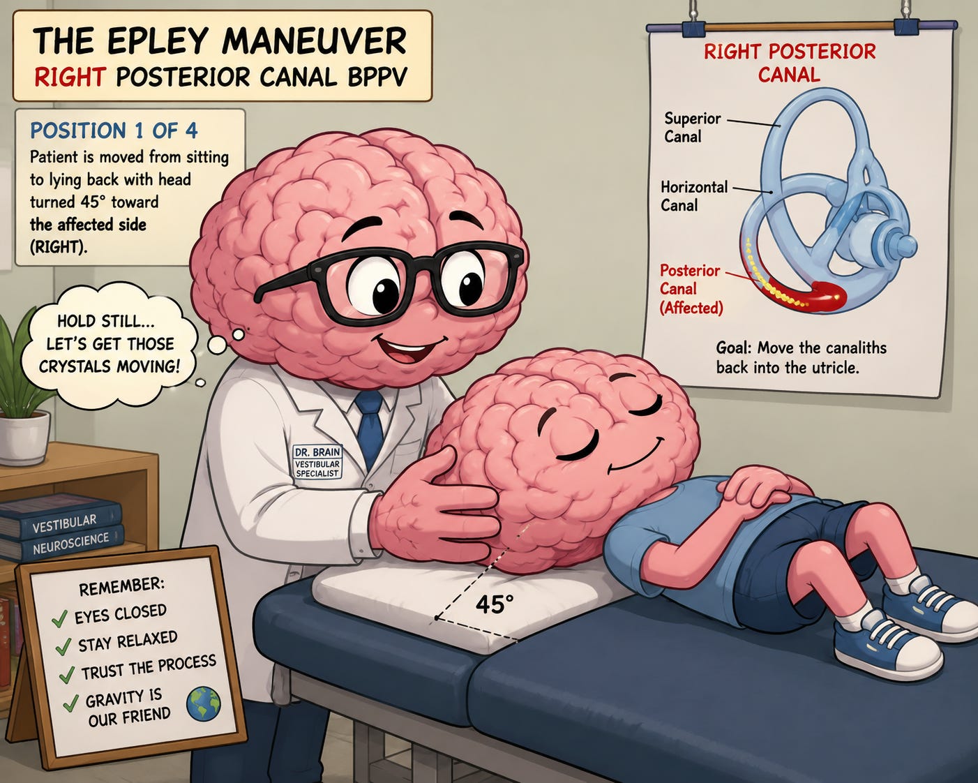
Most clinicians learn that the Epley maneuver, barbecue (BBQ) roll, Semont maneuver, Gufoni maneuver, and other canalith repositioning techniques exist for one purpose: moving displaced otoconia back into the utricle. According to the traditional model, benign paroxysmal positional vertigo (BPPV) is a mechanical disorder. Tiny calcium carbonate crystals leave the utricle, enter one of the semicircular canals, and create abnormal fluid movement whenever the head changes position. Once the crystals are relocated, symptoms improve.
This explanation has helped millions of patients and remains one of the greatest successes in vestibular rehabilitation. Yet it may not represent the entire story. Increasingly, modern neuroscience suggests that every positional maneuver is also a powerful sensory integration exercise for the central nervous system. The maneuver may be repositioning more than crystals—it may also be recalibrating how the brain estimates gravity itself.
The Vestibular System Is Constantly Solving a Physics Problem
Every second of every day, the brain attempts to answer several fundamental questions.
Where is gravity?
Which direction is up?
Am I rotating?
Am I translating?
Is my body tilted?
Am I moving, or is the world moving around me?
These questions sound simple, yet they represent one of the most sophisticated computational problems in neuroscience. The vestibular system continuously combines information from the semicircular canals, otolith organs, neck proprioceptors, joint receptors, muscles, skin, vision, and motor commands to build an internal estimate of body orientation in space. This estimate is never measured directly. Instead, it is inferred from the convergence of multiple sensory systems.
The Brain Does Not Trust Any One Sensory System
The brain functions as a Bayesian prediction machine. Every sensory input receives a level of confidence or weighting. If vision has historically been the most reliable source of information, the brain increases visual weighting. If vestibular information becomes inconsistent, vestibular weighting decreases. If cervical proprioception becomes inaccurate because of injury or chronic muscle tension, neck information becomes less trusted.
The brain continuously adjusts these weightings throughout life. Many chronic dizziness patients develop sensory weighting abnormalities rather than structural vestibular damage. Some become visually dependent. Others become overly dependent on neck proprioception. Others begin ignoring vestibular signals despite having anatomically intact labyrinths. The clinical problem may therefore not simply be damaged receptors but an incorrectly weighted multisensory integration network.
Why Closing the Eyes Changes Everything
One of the most fascinating modifications to positional maneuvers is something incredibly simple: closing the eyes. Vision dominates human orientation. Approximately one-third of the cerebral cortex participates in visual processing in some capacity. When the eyes remain open, the visual system often overrides weaker vestibular information.
Closing the eyes immediately removes the brain’s most dominant orientation signal. Now the nervous system has no choice but to increase reliance upon vestibular information, cervical proprioception, trunk proprioception, pressure receptors in the feet, and internal estimates generated by the cerebellum. This creates an environment where vestibular signals may receive greater central weighting. Rather than allowing vision to solve the orientation problem, the brain must reconstruct gravity using internal sensory information.
Canal-Otolith Convergence
For decades, the semicircular canals and otolith organs were taught as relatively independent systems. Today we understand that they interact continuously. The canals measure angular acceleration. The otoliths measure linear acceleration and gravitational acceleration. Neither system alone can determine body orientation.
The brain must merge both signals to estimate the gravito-inertial acceleration vector. This integration occurs throughout the vestibular nuclei, cerebellar nodulus, uvula, fastigial nucleus, thalamus, and vestibular cortex. Every repositioning maneuver activates canals and otoliths in continuously changing combinations.
As the head rotates, extends, flexes, and rolls, different canal populations become active while the gravitational vector acting upon the otolith organs simultaneously changes. The nervous system is forced to continuously reconcile these changing relationships. Rather than viewing this only as crystal movement, one could also view it as repeated training of canal-otolith convergence.
Holding Each Position Gives the Brain Time to Learn
During traditional repositioning maneuvers, clinicians typically pause for 20 to 60 seconds after each movement. Mechanically, this allows endolymph movement and crystal migration. Neurologically, something equally interesting may be occurring.
While the head remains still, canal activity gradually returns toward baseline while the otolith organs continue signaling head orientation relative to gravity. During this interval, the brain receives a prolonged opportunity to compare changing canal information with stable gravitational information. Repeated exposure may strengthen the internal gravity estimator maintained by the cerebellum. The maneuver becomes not merely a repositioning procedure but a calibration procedure.
Cervical Proprioception Becomes Part of the Therapy
Every head position simultaneously changes cervical muscle length, joint receptor activation, fascial tension, and muscle spindle firing. The upper cervical spine contains one of the highest densities of proprioceptors in the human body. These receptors project extensively into vestibular nuclei and cerebellar structures.
When vision is removed, cervical proprioception becomes increasingly important. Each head position during an Epley or BBQ roll provides a unique combination of vestibular activation and cervical sensory input. The nervous system repeatedly asks whether these sensory signals agree. When they do not, adaptive recalibration may occur.
Patients with cervicogenic dizziness, persistent postural-perceptual dizziness (PPPD), vestibular migraine, concussion, whiplash, and chronic vestibular compensation deficits often demonstrate abnormalities in this integration process.
Downregulating Visual Dependence
Many dizzy patients unknowingly become addicted to vision. Their brains begin relying excessively on visual motion for orientation. This creates difficulty in grocery stores, airports, shopping malls, crowds, scrolling on phones, driving, and visually complex environments. Instead of recalibrating vestibular pathways, the brain compensates by increasing visual gain. Although helpful initially, excessive visual dependence eventually becomes maladaptive.
Performing positional maneuvers with the eyes closed temporarily removes this compensation. The nervous system must solve orientation without visual assistance. Repeated exposure may encourage a more balanced sensory weighting strategy, although direct evidence for this specific application remains limited and should be considered an area for future research rather than established practice.
Gravity Is the Constant Reference Signal
Gravity never turns off. Unlike vision, gravity remains present during darkness. Unlike proprioception, gravity is unaffected by joint position. Unlike hearing, gravity does not depend upon environmental input.
The vestibular system evolved primarily to estimate gravitational orientation. The otolith organs continuously detect the gravitational acceleration vector. The semicircular canals detect changes in head rotation that ultimately modify that gravitational relationship. Every repositioning maneuver repeatedly exposes the nervous system to changing relationships between head orientation and gravity. In many ways, these maneuvers represent structured conversations between the brain and the Earth’s gravitational field.
Beyond BPPV
Could these maneuvers have applications beyond moving otoconia?
This question remains an active area of clinical curiosity. It is biologically plausible that carefully selected positional maneuvers could influence central sensory integration by repeatedly engaging canal, otolith, proprioceptive, and cerebellar networks. However, robust clinical evidence supporting their use specifically as “gravity recalibration” therapies outside of treating BPPV is still lacking.
Patients with chronic vestibular hypofunction, PPPD, vestibular migraine, functional dizziness, concussion, motion sensitivity, space-motion discomfort, visual dependence, and disorders of sensory weighting may benefit from therapies designed to improve multisensory integration. Whether traditional repositioning maneuvers, particularly when modified by eliminating visual input or emphasizing sustained head positions, provide additional benefit beyond established vestibular rehabilitation remains an important research question.
The Cerebellum as the Master Calibrator
The cerebellum functions as one of the brain’s great calibration centers. It continuously compares expected sensory information with actual sensory input. Whenever a mismatch occurs, adaptive learning begins. Every repositioning maneuver creates multiple prediction errors. The canals say one thing. The otoliths say another. The neck provides additional information. The body contributes pressure and proprioceptive feedback. Vision may be absent entirely.
The cerebellum must reconcile all of these inputs into one coherent estimate of orientation. Over time, repeated exposure may strengthen this internal model of gravity and improve confidence in vestibular information.
A Different Way to Think About Positional Therapy
Perhaps repositioning maneuvers should not be viewed solely as mechanical procedures for moving crystals. They may also represent highly organized sensory integration exercises that repeatedly challenge the nervous system to estimate gravity using vestibular, proprioceptive, and cerebellar signals while minimizing visual dominance.
This perspective does not replace the traditional explanation of canalith repositioning; rather, it expands it. The same sequence of head movements that successfully treats BPPV also provides an opportunity for the brain to refine how it integrates information from the semicircular canals, otolith organs, cervical spine, and body into a unified perception of orientation in space.
As our understanding of vestibular neuroscience continues to evolve, future rehabilitation may place greater emphasis not only on repositioning otoconia but also on retraining the brain’s internal estimate of gravity. In that future, the Epley maneuver, the BBQ roll, and similar techniques may be appreciated not only as mechanical treatments for the inner ear, but as sophisticated exercises in multisensory integration that help the nervous system recalibrate one of its most fundamental computations: knowing which way is up.
Translational Therapy: Using Motion to Retrain the Brain’s Perception of Gravity
The Otolith Organs: Your Gravity Sensors
Deep within each inner ear lie two remarkable structures known as the utricle and saccule. Together they form the otolith organs, specialized sensory organs responsible for detecting both gravitational acceleration and linear acceleration.
Unlike the semicircular canals, which detect rotational movements of the head, the otolith organs respond whenever the body accelerates in a straight line. Walking forward, riding in an elevator, stopping suddenly in a car, or stepping sideways all activate the otolith organs.
The challenge is that these organs cannot directly distinguish gravity from acceleration. Both produce force on the sensory hair cells. To the otoliths, tilting your head backward and accelerating forward can create nearly identical sensory signals. This fundamental problem is known as the tilt-translation ambiguity, and solving it is one of the nervous system’s greatest engineering achievements.
The Gravito-Inertial Acceleration Vector
The brain solves this ambiguity by estimating what physiologists call the gravito-inertial acceleration (GIA) vector.
The GIA represents the combination of two forces:
Earth’s gravitational acceleration
Linear inertial acceleration generated by movement
The otolith organs detect only the combined force acting upon them. If you accelerate forward, the inertial component combines with gravity, altering the direction and magnitude of the GIA. Likewise, translating to the left, right, upward, downward, forward, or backward continuously changes the GIA experienced by the vestibular system. The brain then combines this information with signals from the semicircular canals, visual system, neck proprioceptors, muscles, joints, and cerebellum to estimate whether you are moving, tilting, or both.
Gravity Is Not Measured—It Is Estimated
Many people imagine the vestibular system as a simple level or gyroscope. In reality, it behaves more like an advanced navigation computer. Gravity is never measured directly. Instead, the brain continuously predicts where gravity should be based on previous experience and compares those predictions with incoming sensory information. When these predictions become inaccurate, symptoms may emerge.
Patients may describe:
Feeling pulled to one side
Floating sensations
Rocking
Tilting
Walking uphill on flat ground
Feeling as though the floor moves beneath them
Motion intolerance
Spatial disorientation
Chronic dizziness despite normal vestibular testing
In many cases, the issue may not simply be damaged receptors but an internal estimate of gravity that has become maladaptively calibrated.
Using Translation to Challenge the Gravity Estimator
Every linear movement changes the relationship between gravity and inertial acceleration. When performed deliberately, repeated translations expose the nervous system to constantly changing GIA vectors. Imagine slowly translating the entire body toward the right while maintaining an upright head. The otolith organs experience increased rightward inertial acceleration superimposed upon Earth’s downward gravitational pull. The resulting GIA shifts relative to the body.
The cerebellum must determine whether this new sensory signal represents a true body tilt or simply a translation. When repeated many times, this process repeatedly exercises one of the brain’s most important computational tasks: separating gravity from movement. In theory, carefully dosed translation may strengthen the neural networks responsible for estimating gravity.
Every Direction Creates a Different Sensory Problem
Translation is not a single exercise. Each direction creates a unique vestibular computation. Forward translation increases posterior inertial forces relative to the otolith organs. Backward translation produces the opposite effect. Rightward translation shifts the GIA differently than leftward translation. Vertical translations challenge the vestibular system in entirely different ways because they alter the magnitude of the net force acting upon the otolith organs. Diagonal translations combine multiple acceleration vectors simultaneously. Circular translations continuously rotate the GIA relative to the body. Each movement asks the nervous system to solve a slightly different spatial puzzle.
The Brain Learns Through Prediction Errors
The cerebellum is often described as the brain’s master calibration center. Its primary function is not simply movement but learning. Every translation generates predictions. The brain expects a certain vestibular signal based upon motor commands and previous experience. If incoming sensory information differs from that prediction, an error signal is generated. Repeated exposure to these errors drives adaptive plasticity.
Over time, the nervous system refines its internal estimate of gravity, body orientation, and self-motion. This is how healthy vestibular systems remain accurate despite aging, injury, and constantly changing environments.
Sensory Weighting During Translation
Translation also changes how different sensory systems contribute to balance. Closing the eyes removes visual dominance. Standing on compliant surfaces reduces reliable information from the feet. Rotating the head during translation introduces additional canal input. Changing trunk position alters proprioceptive feedback.
The nervous system must determine how much confidence to assign each sensory input. Patients with chronic dizziness often develop abnormal sensory weighting. Some rely excessively on vision. Others become overly dependent upon neck proprioception. Some underutilize vestibular information despite intact peripheral receptors.
Repeated translational exercises may encourage more balanced integration among these sensory systems by forcing the brain to rely more heavily on vestibular and proprioceptive information.
Could Direction Matter?
An intriguing possibility is that not all translations produce identical effects. If one otolith pathway, cerebellar hemisphere, or vestibular network is underperforming, specific directions of translation may preferentially increase activity within those neural circuits.
For example, lateral translations produce asymmetrical activation of the utricular pathways as the brain compares inputs from the two inner ears while interpreting the changing GIA. Likewise, forward-backward and vertical translations engage different populations of otolith afferents and distinct central computations.
Whether individualized directional translation can be used to selectively bias or rehabilitate these networks remains an active area for research, but it provides a compelling framework for personalized vestibular rehabilitation.
Beyond Balance
Gravity perception influences far more than standing upright. The vestibular system contributes to eye movements, posture, spatial memory, navigation, autonomic regulation, emotional processing, and even cognitive function. Vestibular projections extend into the cerebellum, thalamus, hippocampus, insular cortex, parietal cortex, cingulate cortex, basal ganglia, and brainstem autonomic centers.
When gravity estimation becomes distorted, the consequences can extend well beyond dizziness. Patients may experience fatigue, anxiety, visual motion sensitivity, impaired concentration, neck tension, imbalance, persistent motion perception, and reduced confidence during movement. Improving gravity estimation may therefore influence multiple neurological systems simultaneously.
A New Frontier in Vestibular Rehabilitation
Traditional vestibular rehabilitation has focused on gaze stabilization, habituation, balance exercises, and positional maneuvers. These remain highly effective and evidence-based treatments.
Translational therapy offers an additional conceptual framework. Rather than viewing linear movement simply as a way to provoke symptoms or improve tolerance, it can be viewed as a structured method for training the brain’s internal model of gravity by repeatedly manipulating the gravito-inertial acceleration vector.
As our understanding of canal-otolith convergence, cerebellar computation, sensory weighting, and gravity estimation continues to advance, future rehabilitation programs may become increasingly individualized. Carefully selected translational movements—performed in specific directions, at specific speeds, and under controlled sensory conditions—could one day become an important tool for improving the brain’s perception of orientation in space.
Ultimately, rehabilitation may prove to be less about treating dizziness itself and more about restoring one of the brain’s most fundamental abilities: accurately knowing where the body exists within Earth’s gravitational field.
REFERENCES:
Angelaki DE, Cullen KE. Vestibular system: The many facets of a multimodal sense. Annu Rev Neurosci. 2008;31:125-150.
Goldberg JM, Wilson VJ, Cullen KE, Angelaki DE, Broussard DM, Buttner-Ennever JA, Fukushima K, Minor LB. The Vestibular System: A Sixth Sense. New York, NY: Oxford University Press; 2012.
Laurens J, Angelaki DE. The functional significance of velocity storage and its dependence on gravity. Exp Brain Res. 2011;210(3-4):407-422.
Oman CM. A heuristic mathematical model for sensory conflict and motion sickness. Acta Otolaryngol Suppl. 1982;392:4-44.
Yakusheva TA, Blazquez PM, Angelaki DE. Relationship between complex and simple spike activity in macaque caudal vermis during three-dimensional vestibular stimulation. J Neurosci. 2010;30(24):8111-8126.
I am not deeply knowledgeable about the brain and body but found this article fascinating as to the hidden connections we take for granted as they orient us spatially.
The intricacies of evolution you have outlined leaves awe. Gravity, the inner ear, all the interwoven aspects of human anatomy allowing functioning.
Why not perform maneuvers standing? Gravity effects the body in this position the most.