
Transcranial Photobiomodulation: Can Light Improve Brain Function?
The Emergence of Transcranial Photobiomodulation

By Dr. David Traster, DC, MS, DACNB
Co-owner, The Neurologic Wellness Institute
Boca Raton • Chicago • Waukesha • Wood Dale
www.neurologicwellnessinstitute.com
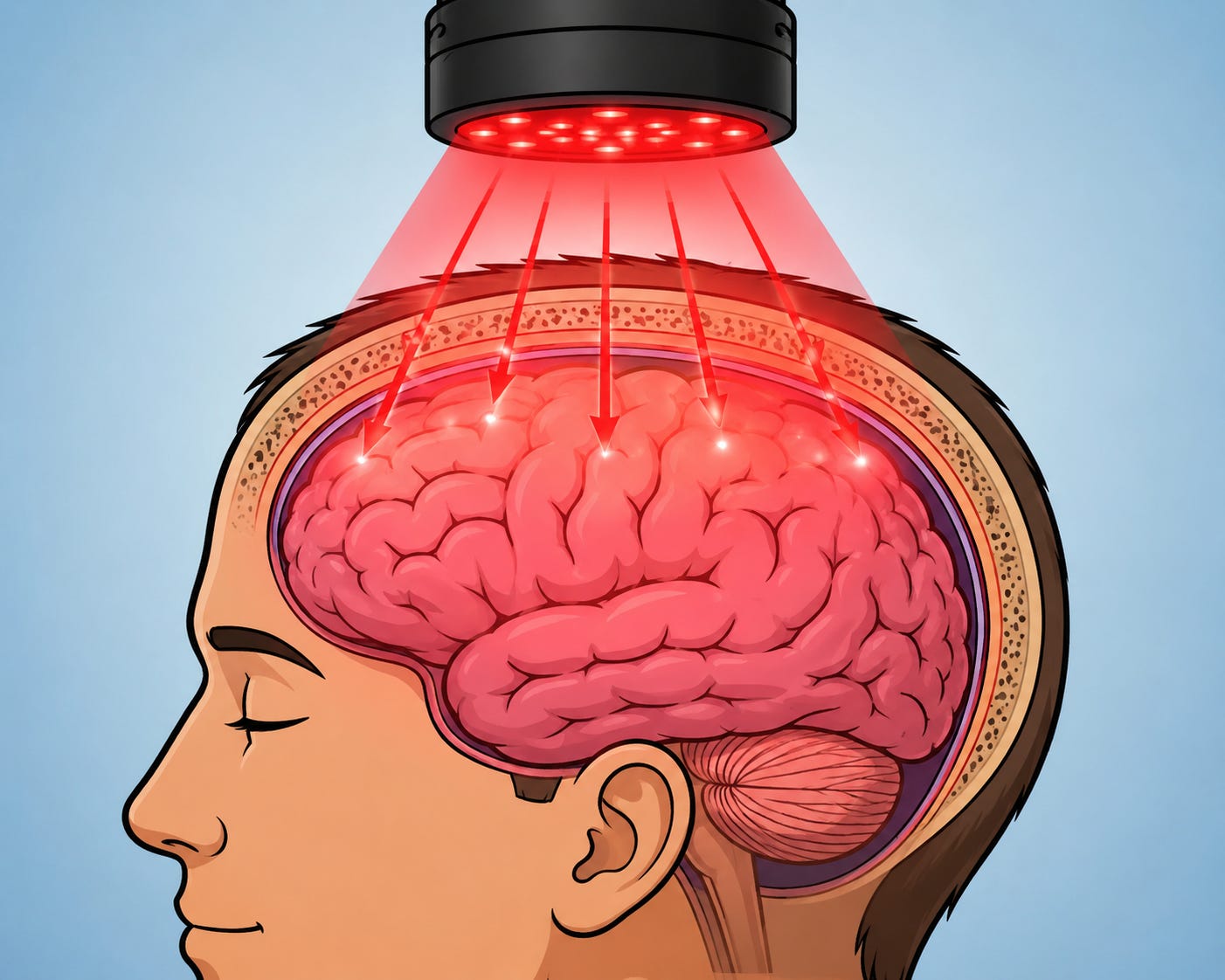
Transcranial photobiomodulation (tPBM), also referred to as transcranial low-level light therapy, is an emerging field of neuroscience that investigates whether specific wavelengths of light can penetrate the scalp and skull to influence brain physiology. Unlike surgical procedures or implanted neurostimulation devices, transcranial photobiomodulation is entirely noninvasive. The therapy typically utilizes red and near-infrared light delivered through lasers or high-powered LEDs to expose the scalp over selected cortical regions.
Although light has been used therapeutically for centuries, modern photobiomodulation began to develop after Hungarian physician Endre Mester unexpectedly observed accelerated wound healing in the 1960s while investigating whether low-powered lasers caused cancer. Instead, low-energy laser exposure appeared to stimulate tissue repair. Over subsequent decades, researchers recognized that these biological effects extended well beyond skin and muscle into neural tissue.
Only during the past twenty years has substantial attention focused on whether sufficient light can penetrate the human skull to directly influence cortical neurons, cerebral blood vessels, glial cells, and mitochondrial metabolism. Today, research is exploring applications ranging from traumatic brain injury and stroke to Alzheimer’s disease, depression, Parkinson’s disease, cognitive enhancement, post-concussion syndrome, and neurorehabilitation.
How Does Photobiomodulation Work?
Unlike high-powered surgical lasers that cut or destroy tissue through heat, photobiomodulation operates through photochemical mechanisms rather than thermal injury.
The primary chromophore believed to absorb red and near-infrared light inside cells is cytochrome c oxidase, also known as Complex IV of the mitochondrial electron transport chain. Absorption of photons appears capable of temporarily improving mitochondrial respiration and ATP production while simultaneously dissociating nitric oxide from cytochrome c oxidase, allowing oxygen utilization to improve.
The downstream physiological effects described in laboratory studies include:
Increased ATP production
Improved mitochondrial membrane potential
Increased cerebral blood flow
Nitric oxide release
Reduced oxidative stress
Modulation of inflammatory cytokines
Increased neurotrophic factors including BDNF
Enhanced synaptic plasticity
Promotion of neurogenesis in animal models
Improved lymphatic and glymphatic clearance in preliminary studies
Rather than stimulating one isolated brain region, photobiomodulation appears to influence the metabolic health of neurons and glial cells themselves.
The Optical Window of the Human Brain
Not every wavelength of light penetrates biological tissue equally.
Human tissue contains multiple substances that absorb light:
Melanin
Hemoglobin
Water
Lipids
Bone minerals
These molecules determine how deeply photons travel.
Researchers commonly refer to the “optical window” of biological tissue, generally ranging from approximately 650 to 1100 nanometers (nm). Within this range, absorption by hemoglobin decreases while absorption by water remains relatively low, allowing deeper tissue penetration.
This explains why nearly all modern transcranial photobiomodulation research focuses on red and near-infrared wavelengths.
Common Wavelengths Used in Research
630–660 nm (Red Light)
Red light represents one of the earliest wavelengths studied.
Potential effects include:
Increased superficial blood flow
Enhanced wound healing
Reduced inflammation
Improved scalp circulation
Mild cortical penetration
Estimated penetration:
Scalp: excellent
Skull: moderate
Cortex: approximately 5–15 mm depending on anatomy and optical power
Because skull bone absorbs a significant portion of visible red light, only a small percentage ultimately reaches cortical tissue.
670 nm
670 nm has received considerable attention because laboratory studies suggest particularly efficient absorption by cytochrome c oxidase.
Animal studies have demonstrated:
Improved retinal function
Reduced inflammation
Neuroprotection
Mitochondrial enhancement
Human brain studies remain relatively limited but promising.
808–810 nm
This is arguably the most extensively studied wavelength for transcranial photobiomodulation.
Numerous clinical investigations have reported improvements in:
Working memory
Executive function
Attention
Depression symptoms
Chronic traumatic brain injury
Cerebral oxygenation
Functional MRI connectivity
Because 810 nm penetrates tissue efficiently while maintaining strong mitochondrial absorption, many investigators consider it the current “sweet spot” for brain applications.
Estimated cortical penetration:
Approximately 20–40 mm beneath the scalp under favorable conditions.
830 nm
830 nm shares many characteristics with 810 nm.
Research suggests:
Enhanced ATP production
Increased cerebral blood flow
Reduced neuroinflammation
Improved neuronal survival
Many commercially available photobiomodulation helmets utilize 810 and 830 nm simultaneously.
850 nm
850 nm LEDs are among the most widely marketed consumer devices.
Advantages include:
Good tissue penetration
Less expensive LEDs
Lower manufacturing costs
Research suggests physiological activity, although cytochrome c oxidase absorption may not be quite as efficient as 810 nm.
904 nm
904 nm pulsed lasers have historically been used in musculoskeletal rehabilitation.
Some neurological investigations have explored their use because deeper penetration may occur under certain conditions.
980 nm
980 nm penetrates deeply but water absorption begins increasing substantially.
This creates greater heat production, making it less commonly used for brain photobiomodulation.
1064 nm
1064 nm has become increasingly popular in university research.
Multiple studies have demonstrated:
Increased cerebral oxygenation
Improved executive function
Enhanced attention
Increased prefrontal metabolism
Because scattering decreases as wavelength increases, 1064 nm may penetrate somewhat deeper than shorter wavelengths despite reduced mitochondrial absorption.
What About Other Colors?
Blue Light (450–495 nm)
Blue light penetrates poorly into the brain.
Research primarily focuses on:
Circadian rhythm regulation
Melatonin suppression
Retinal ganglion cells
Mood disorders
Blue wavelengths generally do not reach cortical tissue in meaningful amounts through the intact skull.
Green Light (520–550 nm)
Green light has been investigated for:
Migraine
Pain modulation
Visual comfort
Retinal physiology
Very little reaches the cortex.
Yellow and Orange Light
Minimal evidence supports transcranial neurological applications.
Most effects remain superficial.
Violet Light (400–430 nm)
Violet light penetrates the least.
Applications include:
Antimicrobial therapy
Dermatology
Surface wound healing
It has essentially no meaningful cortical penetration.
How Deep Does Light Actually Reach?
Penetration is influenced by:
Hair thickness
Skin pigmentation
Skull thickness
Frontal versus temporal bone
Cerebrospinal fluid
Brain blood flow
Age
Optical power
Beam collimation
Only a small percentage of emitted photons reach cortical tissue—often estimated at less than 5%, and in many experimental conditions substantially less. Nevertheless, animal and human studies suggest that even this small photon flux may be sufficient to produce measurable biological effects, indicating that absolute energy at the target may be more important than transmission percentage alone.
Living Skull vs. Cadaver Skull Research
This distinction is often overlooked but critically important.
Many early optical penetration studies used cadaver skulls because they were easier to obtain and allowed direct optical measurements.
However, cadaver skulls differ from living skulls in several important ways:
Loss of blood circulation
Reduced tissue hydration
Changes in cerebrospinal fluid dynamics
Altered optical scattering
Different refractive indices
Absence of pulsatile blood flow
Living tissue contains continuously moving blood and highly organized water molecules that influence photon propagation through absorption and scattering. Cerebrospinal fluid is relatively transparent in the red and near-infrared spectrum and can facilitate photon transmission compared with dense bone, but the overall effect depends on geometry and the complex optical properties of surrounding tissues. Consequently, penetration measurements from cadaver experiments should not be assumed to directly represent photon delivery in living humans.
Modern studies increasingly combine computational modeling with measurements in living participants using techniques such as near-infrared spectroscopy (NIRS), magnetic resonance imaging (MRI), and diffuse optical tomography to estimate intracranial light distribution more realistically.
Does Pulsing Matter?
One of the most debated questions in photobiomodulation concerns whether continuous-wave light or pulsed light produces greater biological effects.
Several hypotheses have been proposed.
Pulsing may:
Reduce tissue heating
Improve photon efficiency
Better synchronize with neuronal oscillations
Reduce receptor desensitization
Enhance mitochondrial signaling
Some animal and early human studies suggest pulsed delivery can produce stronger biological responses than continuous illumination at the same average energy, but results are not uniform across wavelengths, doses, or disease models. At present, no single pulse frequency has been established as superior for all neurological applications.
Brain-Wave Frequencies
Several pulse frequencies have attracted research interest because they overlap with endogenous neural oscillations.
10 Hz (Alpha)
Investigated for:
Relaxation
Anxiety
Attention
Some studies suggest improvements in cognitive performance.
40 Hz (Gamma)
One of the hottest areas of neuroscience.
Gamma-frequency sensory stimulation has been investigated for:
Alzheimer’s disease
Memory
Microglial activation
Amyloid clearance
Functional connectivity
Most of this literature involves visual or auditory flicker rather than transcranial laser stimulation, but some photobiomodulation systems also employ 40 Hz pulsing as an experimental approach.
Theta Frequencies (4–8 Hz)
Explored for:
Memory
Learning
Hippocampal function
Evidence remains preliminary.
Delta Frequencies
Occasionally explored in sleep research, though evidence for therapeutic brain photobiomodulation is limited.
What About Rife Frequencies?
Some commercial devices advertise “Rife frequencies” based on the early twentieth-century ideas of Royal Raymond Rife, proposing that specific electromagnetic frequencies can selectively destroy pathogens or treat disease.
At present, there is no high-quality clinical evidence demonstrating that applying Rife frequencies through transcranial photobiomodulation improves neurological disorders. These claims remain outside the mainstream evidence base, and they should not be considered established mechanisms of photobiomodulation.
LED Versus Laser
This remains one of the most common clinical questions.
Lasers
Advantages:
Coherent light
Collimated beam
Greater power density
Better penetration
More precise targeting
Disadvantages:
Higher cost
Greater eye hazard
Smaller treatment area
More operator training
LEDs
Advantages:
Much safer
Less expensive
Larger treatment area
Easier home use
Lower eye risk
Disadvantages:
Less collimated light
Greater beam divergence
Lower irradiance at depth
Importantly, once photons enter tissue, coherence is rapidly lost because biological tissues scatter light extensively. This has led many researchers to conclude that wavelength, irradiance, fluence, and total energy delivered are more important determinants of biological effect than coherence alone. Well-designed LED systems have demonstrated clinical benefits in several studies despite lacking coherent laser output.
Laser Classifications: Class 1 Through Class 4
Laser classes describe hazard potential, not therapeutic effectiveness.
Class 1
These lasers are considered safe during normal operation because accessible emissions remain below hazardous exposure limits.
Advantages include an excellent safety profile, but their low output generally makes them impractical for transcranial photobiomodulation unless many emitters are combined in a specialized device.
Class 2
Class 2 lasers emit visible light up to approximately 1 mW and rely on the normal blink reflex for eye protection.
They are primarily used as alignment or pointing devices rather than therapeutic tools and very few class 2 devices have been used for brain photobiomodulation outside of companies self-funded research.
Class 3
Class 3 is divided into:
Class 3R (formerly IIIa)
Class 3B (formerly IIIb)
Class 3B therapeutic lasers are among the most commonly studied systems in photobiomodulation research. They provide sufficient irradiance to deliver therapeutic doses to cortical tissue while maintaining a lower thermal risk than surgical lasers. Appropriate eye protection remains essential.
Class 4
Class 4 lasers exceed 500 mW and can rapidly heat tissue if misused.
Potential advantages include:
Higher photon delivery
Shorter treatment times
Greater ability to treat larger areas
Potential disadvantages include:
Increased risk of thermal injury
Greater eye hazards
Need for careful dosing and movement to avoid overheating
Although some clinicians use Class 4 devices for transcranial applications, most published brain photobiomodulation studies have employed lower-power systems designed to deliver nonthermal doses. Current evidence does not show that simply using a higher laser class leads to better neurological outcomes; appropriate wavelength, irradiance, fluence, exposure time, and total energy appear to be more important than maximum power alone.
Clinical Applications Under Investigation
Research continues to expand into numerous neurological conditions, including:
Mild traumatic brain injury
Persistent post-concussion symptoms
Alzheimer’s disease
Mild cognitive impairment
Parkinson’s disease
Major depressive disorder
Anxiety disorders
Stroke recovery
Multiple sclerosis
Chronic fatigue syndrome
Long COVID-related cognitive dysfunction
Migraine
Healthy cognitive enhancement
Evidence is strongest for improving cerebral blood flow, cerebral oxygen metabolism, and selected cognitive outcomes, while studies in neurodegenerative diseases remain encouraging but preliminary. Many trials are small, protocols vary considerably, and larger randomized controlled studies are needed before firm clinical recommendations can be made.
The Future of Brain Photobiomodulation
Transcranial photobiomodulation represents one of the most intriguing developments in noninvasive neuroscience. Rather than forcing neurons to fire, it aims to improve the metabolic environment in which neurons operate by supporting mitochondrial function, vascular physiology, and cellular resilience. The field has progressed from basic laboratory observations to human clinical trials demonstrating changes in cerebral oxygenation, functional connectivity, cognition, and mood. However, important questions remain regarding the optimal wavelength, dose, pulse structure, treatment duration, and patient selection.
Current evidence most strongly supports the use of red and near-infrared wavelengths—particularly around 670 nm, 810 nm, 830 nm, 850 nm, and 1064 nm—for transcranial applications because these wavelengths penetrate tissue more effectively than shorter visible wavelengths and interact with known biological targets. At the same time, claims involving proprietary pulse frequencies, Rife frequencies, or dramatically superior performance of any single device should be viewed cautiously until supported by rigorous clinical trials. As imaging technologies, dosimetry, and optical modeling continue to improve, transcranial photobiomodulation may become an increasingly important adjunctive tool in neurorehabilitation, cognitive medicine, and the treatment of neurological disease.
References
Hamblin, M. R. (2016). Mechanisms and applications of the anti-inflammatory effects of photobiomodulation. AIMS Biophysics, 3(3), 337–361.
Hamblin, M. R. (2018). Photobiomodulation for traumatic brain injury and stroke. Journal of Neuroscience Research, 96(4), 731–743.
Salehpour, F., Mahmoudi, J., Kamari, F., Sadigh-Eteghad, S., Rasta, S. H., & Hamblin, M. R. (2018). Brain photobiomodulation therapy: A narrative review. Molecular Neurobiology, 55(8), 6601–6636.
Cassano, P., Petrie, S. R., Mischoulon, D., Cusin, C., Henderson, T. A., Lee, C. K., ... & Hamblin, M. R. (2016). Review of transcranial photobiomodulation for major depressive disorder. Neurophotonics, 3(3), 031404.
Naeser, M. A., Ho, M. D., Martin, P. I., Hamblin, M. R., Koo, B. B., & Pascual-Leone, A. (2023). Transcranial photobiomodulation: Current evidence and future directions for neurological rehabilitation. NeuroRehabilitation, 52(3), 281–299.
So insughtful
Are there models about what the photons actually do at the molecular level? E.g. how they enlarge the ATP-availability?